What Causes an Inverted T-Wave?
Definition
The T wave is the ECG manifestation of ventricular repolarization of the cardiac electrical cycle. The T wave is normally upright in leads I, II, and V3 to V6; inverted in lead aVR; and variable in leads III, aVL, aVF, V1, and V2. Thus, T-wave inversions in leads V1 and V2 may be fully normal.
Causes of Inverted T-Waves
A variety of clinical syndromes can cause T-wave inversions; these range from life-threatening events, such as acute coronary ischemia, pulmonary embolism, and CNS injury.
Primary and secondary t wave inversions- The causes of T-wave inversions have commonly been grouped into 2 categories: primary T-wave changes and secondary T-wave changes. Alterations in the duration or morphology of the action potential, without concurrent changes in the orderly sequence of activation, are termed “primary changes.” Primary T-wave inversions are associated with benign syndromes, such as the persistent juvenile T-wave pattern and the digitalis effect, as well as morbid conditions, including acute coronary ischemic events and CNS catastrophe. Secondary T-wave changes result from aberrant ventricular activation in the context of normal action potential characteristics. Examples include bundle branch blocks, preexcitation states, and ventricular ectopic or paced beats.
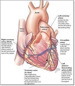
Coronary artery disease- T-wave inversions associated with coronary artery disease may result from myocardial ischemia, non–ST-segment elevation acute myocardial infarction (MI) (Figure), or previous MI. In addition, an important subgroup of patients with unstable angina present with significantly abnormal T-wave inversions—either symmetric, deeply inverted T waves or biphasic T waves in the precordial leads (V2 and V3 in particular). Patients with this history and these ECG findings have Wellen syndrome, which is frequently associated with proximal left anterior descending coronary artery critical stenosis.
Neurogenic causes- There are a number of neurogenic causes of primary T-wave inversions, each of which has characteristic features. For example, T waves in patients who have sustained a cerebrovascular accident (CVA) have a distinctly deep, widely splayed appearance with an outward bulge of the ascending limb that results in a striking asymmetry. This has been termed the “CVA T-wave pattern.” CNS hemorrhage and status epilepticus are also associated with T-wave abnormalities.
Pulmonary causes- Patients with pulmonary embolism (PE) may also display T-wave abnormalities, including T-wave inversions. The T-wave findings in these patients are typically shallow inversions in the inferior leads. Deeper T-wave inversions— attributed to acute right ventricular strain and occasionally seen in patients with massive PE—are generally observed in the right to mid precordial leads.
Arrhythmias- Right bundle branch block, with its associated depolarization- repolarization changes, may produce marked ST-segment–T-wave changes.